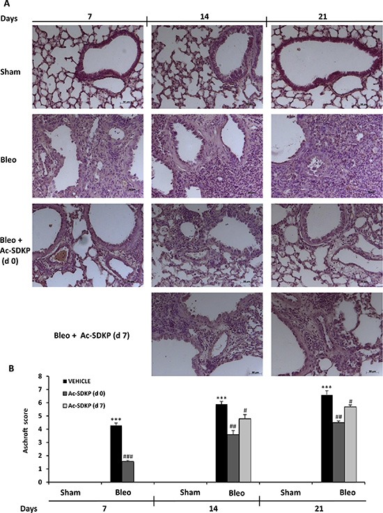
Figure 3. Ac-SDKP treatment suppressed BLEO-induced histological marks of lung damage and fibrosis in mouse lung.

(A) Representative microphotographs (150 ×)of FFPE lung tissue slices stained by H & E from indicated mice at the indicated time points. (B) Semi-quantitative fibrosis scoring was assessed according to the Ashcroft scale. Slides were scored by a single investigator in a blinded fashion. Graphs show mean values ± s.d. of at least five mice for each group. *P < 0,05 vs Sham; **P < 0,01 vs Sham; ***P < 0,001 vs Sham; #P < 0,05 vs Bleo; ##P < 0,01 vs Bleomycin; ###P < 0,001 vs Bleo.
