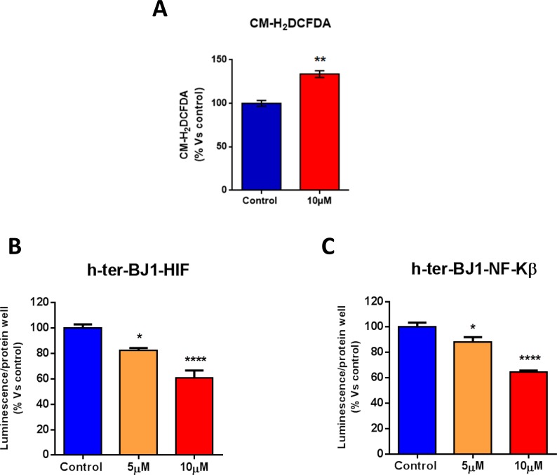
Figure 7. Atovaquone treatment does not induce pro-inflammatory or stress-induced responses in human normal fibroblasts.
A. ROS levels were measured in normal human fibroblasts treated with atovaquone (10 μM) or DMSO for 48 hours. Note that atovaquone increases ROS levels. B., C. The activation of HIF and NFκB pathways was monitored using Luc-reporter fibroblasts carrying HIF- and NFκB-Luc reporter elements. Luciferase assays were performed on cells treated with atovaquone (5μM and 10 μM) or DMSO control for 48 hours. Luminescence was normalized using SRB (total proteins), as a measure of cell viability. Note that atovaquone treatment decreases the activation of HIF (B) and NFκB (C) pathways in normal fibroblasts, in a dose-dependent manner. *p < 0.01, **p < 0.001, ****p < 0.00001 evaluated with one-way ANOVA.

