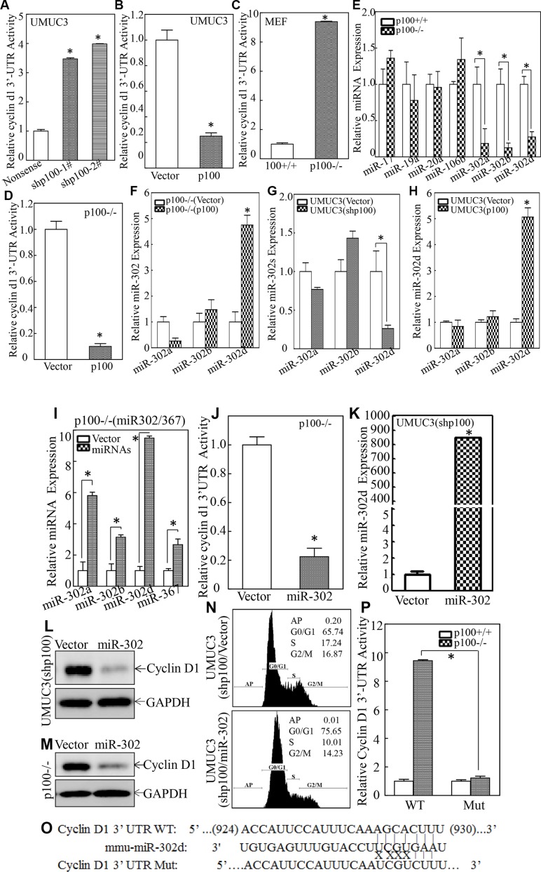
Figure 4. miR-302d was specific activated by p100 and directly inhibited Cyclin D1 protein translation.
(A–D) cyclin d1 3′UTR luciferase reporter was transiently transfected into the indicated cells and luciferase activity of each transfectant was evaluated. The results were presented as relative cyclin d1 3′-UTR activity. The symbol (*) indicates a significant difference (p < 0.05). (E–H) The levels of indicated microRNAs were evaluated by quantitative real-time PCR. The symbol (*) indicates a significant difference as compared with control cells as indicated (p < 0.05). (I) p100−/− cells were stably transfected with construct of miR-302/367 and the miRNA expression levels were determined by real-time PCR. The symbol (*) indicates a significant increase in comparison to the scramble control transfectant (p < 0.05). (J) The cyclin d1 3′-UTR luciferase reporter was transiently transfected into p100−/−(Vector) or p100(miR-302) cells. Luciferase activity of each transfectant was evaluated and the results were presented as relative cyclin d1 3′-UTR activity. The symbol (*) indicates a significant decrease as compared with that in vector transfectant (p < 0.05). (K) UMUC3(shp100) cells were stably transfected with construct of miR-302/367. The miR-302d expression was determined by real-time PCR and the symbol (*) indicates a significant increase as compared with control vector transfectant (p < 0.05). (L and M) The cell extracts as indicated were subjected to Western Blot and GAPDH was used as a protein loading control. (N) Flow-cytometry analysis of cell cycle alteration was carried out as indicated. (O) Schematic of the construction of the cyclin d1 mRNA 3′-UTR luciferase reporter and its mutants were aligned with miR-302d. (P) Wild-type and mutant of cyclin d1 3′-UTR luciferase reporters were co-transfected with pRL-TK into p100+/+ and p100−/− cells, respectively. Luciferase activity of each transfectant was evaluated and the results were presented as relative cyclin d1 3′-UTR activity. The symbol (*) indicates a significant decrease in cyclin d1 3′-UTR activity as compared with that in WT cyclin d1 3′-UTR reporter transfectant (p < 0.05).

