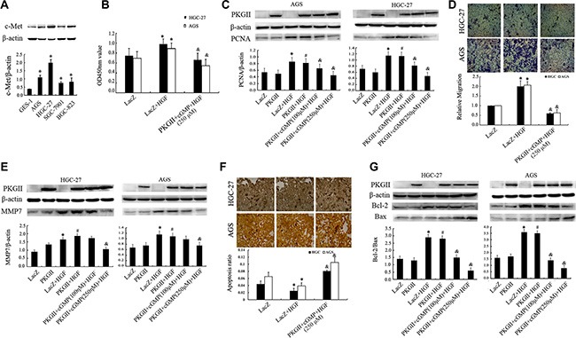
Figure 1. Analysis of the effect of PKG II on HGF-induced changes of proliferation, migration and apoptosis activities.

(A) The expression of c-Met in human gastric epithelial cell line GES-1 and gastric cancer cell lines AGS, HGC-27, SGC-7901 and BGC-823. (*P < 0.05, compared to GES-1 cell). (B–G) The detection of proliferation, migration, apoptosis activities and related protein expression. The AGS and HGC-27 cells were infected with either Ad-LacZ or Ad-PKG II, serum-starved for 12 h, stimulated with 8-pCPT-cGMP (100 or 250 μΜ) for 1 h, and then treated with HGF (50 ng/ml) for 24–48 h. (B) The relative proliferation activities were presented. (C) The expression of PCNA was detected by Western blotting with an anti-PCNA antibody. (D) The transwell migration assay was applied to detect the migration activity. The representative data showed the relative activities of migration. (E) The expression of MMP7 was detected by Western blotting with an anti-MMP7 antibody. (F) TUNEL method was applied to analyze the apoptosis activity, and the average ratio of apoptotic cells per field (magnification, × 200) was shown. (G) Detection of Bax and Bcl-2 protein by Western blotting in different groups. (*P < 0.05, compared to LacZ group; #P < 0.05, compared to PKG II group; &P < 0.05, compared to LacZ+HGF group, or PKG II+HGF group).
