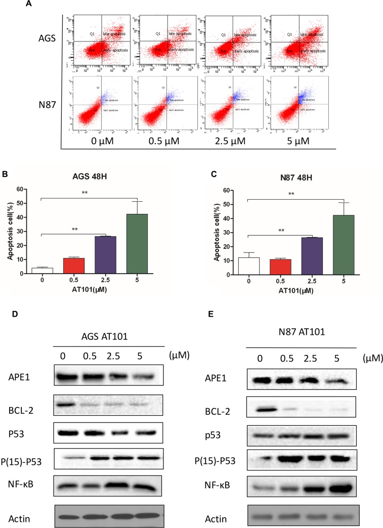
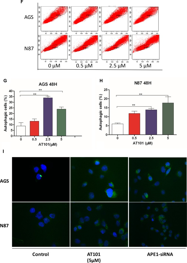
Figure 3. Inhibition of APE1 by AT101 promotes apoptosis and autophagy of gastric cancer cells.
AGS and NCI-N87 cells treated with AT101 at different concentrations of 0.5, 2.5, and 5 μM for 48 hours. (A–C) The apoptosis of cells was quantitated on the graph using the Annexin V: PE apoptosis detection kit and a flow cytometer. (t test; *, P < 0.05) (D, E) The markers of BCL-2, P53, Phosphate-activated P53 (Ser15) and NF-κB were detected by western blot assay. (F–H) The autophagic cells was detected by the Cyto-IDr autophagy detection kit and analyzed using the green (FL1) channel of the flow cytometer. (I) The cellular autophagy induced by the concentration of AT101 (5 μM) and APE1 siRNA was examined by confocal microscopy with the application of Cyto-IDr autophagy detection kit.


