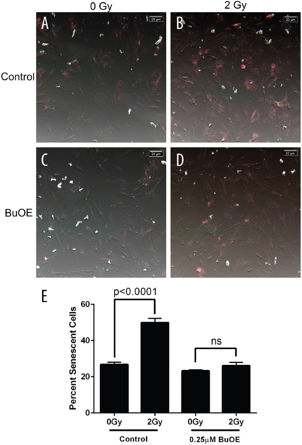
Figure 3. Radiation-induced senescence is reduced in primary colorectal fibroblasts grown in MnTnBuOE-2-PyP.

Mouse colorectal fibroblasts maintained in media with PBS (Control) or 0.25 μM MnTnBuOE-2-PyP (BuOE) were either sham irradiated A. & C. or treated with 2 Gy x-rays B & D., 48 hours later cells were stained for SA-β-gal expression. Cells were imaged with an inverted brightfield microscope, these images were inverted (A-D) to aid in counting SA-β-gal positive (pink) cells. E. Quantification of senescent cells. SA-β-gal expression is significantly increased (p<0.0001) in control cells receiving 2 Gy as compared to the unirradiated population. When cells were grown in BuOE there was no significant increase (p=0.22) in SA-β-gal expression following radiation treatment. All data are representative of the mean and standard deviation and were obtained from 3 independent experiments. The differences of mean percent senescent cells were analyzed for significance using 1-way ANOVA followed by post hoc Tukey's test for multiple comparisons.
