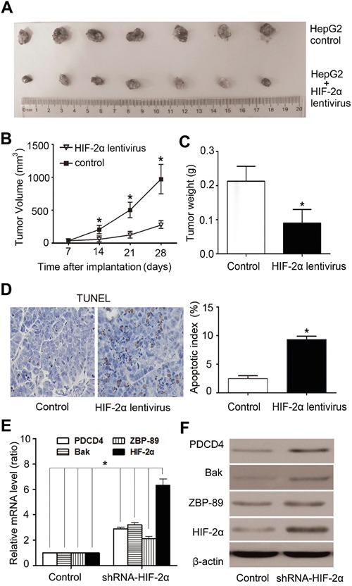
Figure 6. Over-expression of HIF-2α induced HCC growth arrest and high apoptosis rate in HepG2-cell xenografts in nude mice.

A. Tumors formed by implanted cells with different expression levels of HIF-2α. B. Tumor volume. Tumor volumes were calculated and data were plotted using the geometric mean for each group vs. time. Each point represents the mean tumor volume (± sd) of measurements from the 7 mice in each treatment group. **P<0.01, two-tailed test. C. The weight of tumors was significantly lower in HIF-2α lentivirus-infected group than the control group. **P<0.01, two-tailed test. D. Cells positive for TUNEL staining were statistically increased in HIF-2α-over-expressing tumors, compared with the control (**P < 0.01, Student's t test). Real-time PCR E. and western blotting analysis F. of HIF-2α, ZBP-89, Bak, PDCD4 expression in xenograft tumors after mouse sacrifice.
