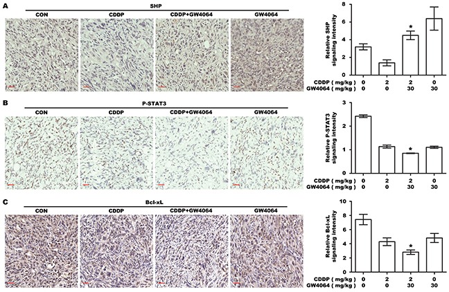
Figure 7. The effect of FXR agonist/CDDP co-treatment on SHP-STAT3-Bcl-xL signaling in vivo.

A. IHC staining for the expression of SHP in transplanted tumor tissues. The nucleus and cytoplasm brown staining represented positive labeling for SHP. Scale bar: 30 μm. The chart was the quantification of SHP in xenografted tumors. *P <0.05, combination treatment group compared with CDDP-alone group. B. IHC staining for P-STAT3. The nucleus brown staining represented positive signal for P-STAT3. Scale bar: 30 μm. The chart was the quantification. *P <0.05, combination treatment group compared with CDDP-alone group. C. IHC staining for Bcl-xL. The cytoplasm brown staining represented positive labeling for Bcl-xL, and the nucleus was stained by hematoxylin. Scale bar: 30 μm. The chart was the quantification. *P <0.05, combination treatment group compared with CDDP-alone group.
