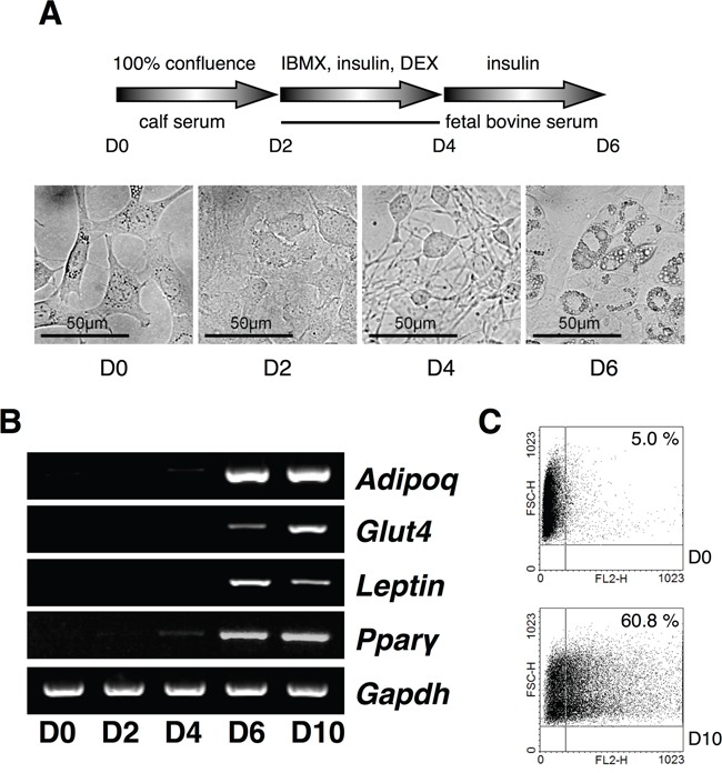
Figure 1. Expression analysis of USPs in adipocyte differentiation.

A. A scheme for the induction of adipocytes with IBMX, DEX, and insulin from 3T3-L1 cells, and the cell morphology was checked every a couple of days at original magnification 40×. B. Primers for Adipoq, Glut4, Leptin, and Ppar-γ were used for RT-PCR using cDNA from each phase of the differentiating adipocytes. C. The differentiated adipocytes were assorted and analyzed by fluorescence-activated cell sorting (FACS).
