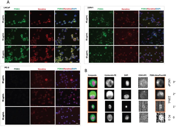
Figure 3.

A. Immunostaining for PSMA and pan-keratin comparing different prostate cancer cell line cells (LNCap, 22Rv1, and PC-3). DAPI was used for nuclear counterstainings. Different concentrations of the PSMA BioLegend antibody (clone LNI-17) were applied (40 - 60 μg / mL). B. Detection of PSMA expression on tumor cells (LNCaP, 22Rv1, and PC-3) spiked into 7.5 mL of healthy donors blood by the CellSearch® system.
