
Figure 3. Metastasis suppressive effects of miR-199b in CRC cell lines in vitro and in vivo.
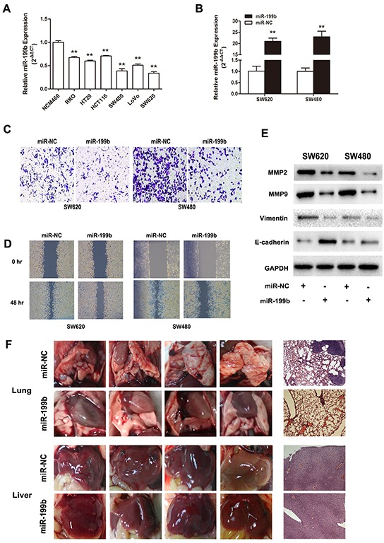
A. Relative miR-199b expression level in six CRC cell lines compared to the normal colorectal cell line NCM460. The average gene expression from NCM460 was designated as 1. B. Relative expression of miR-199b after transfected with miR-199b mimics and its negative control (NC), detected by qRT-PCR. The average miRNA expression from NC was designated as 1. C. The invasive ability of SW480 and SW620 cells was assessed by Transwell assay after overexpression of miR-199b. Statistics analysis was performed by counting the stained cells that invaded to the lower chamber under a light microscopy. D. Wound healing assay was applied to measure the migration ability of SW620 and SW480 cells. Quantification was performed by measuring the smallest clearance distance of the wound. E. Western blot analysis showed the expression levels of invasion related molecules MMP2 and MMP9, the epithelial-mesenchymal transition (EMT) marker E-cadherin and Vimentin after overexpression of miR-199b. F. Pictures of the lungs and livers in nude mice and their respective representative images of the tissues by hematoxylin-eosin (HE) staining. Metastases in lungs were observed in NC group but little in miR-199b group. No obvious metastasis formation could be seen in liver in these two groups. *P<0.05, **P<0.01.