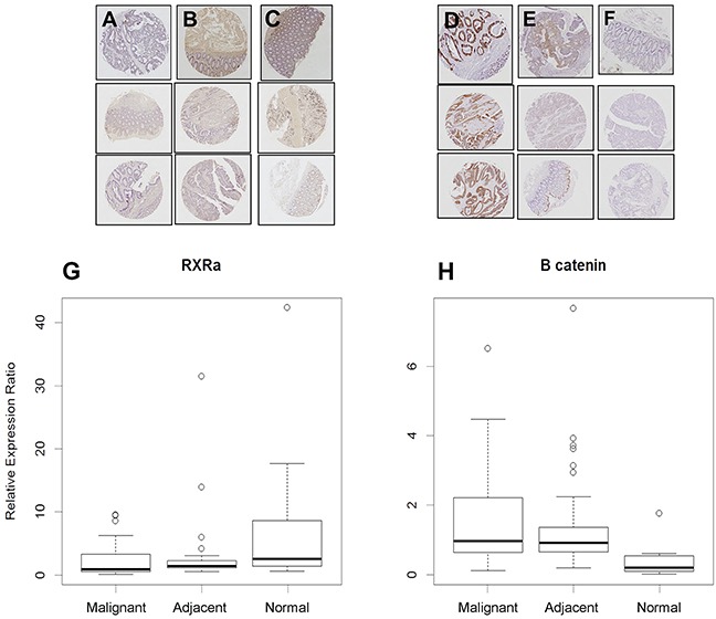
Figure 1. RXRα and β-catenin immunostaining in human tissues.

A. RXRα staining is notably absent in human colon tumors, but present in B. adjacent and C. normal colon mucosa (*p-values: normal vs. adjacent = 0.029, normal vs malignant = 0.0035, adjacent vs malignant = 0.23) while D. β-catenin staining is high in human colon tumors and in E. adjacent tissues but low in F. normal colon mucosa. G. Quantitated expression of RXRα and H. β-catenin in various tissues, data displayed as box and whisker plots (black line – mean, circles outliers, box 50% mean distribution and lines 99% distribution) (*p-values: normal vs adjacent = 0.001, normal vs malignant = 0.0005, adjacent vs malignant = 0.48).
