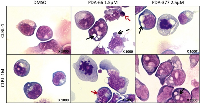
Figure 3. The morphological changes after PDA-66 and PDA-377 treatment.

CLBL-1 and CLBL-1M cells were incubated with 1.5 μM PDA-66, 2.5 μM PDA-377 and DMSO. Cytospins of treated cells were stained with Pappenheim method after 72 h. Representative pictures are displayed. In DMSO controls, lymphoid round cells show moderately abundant light to dark blue cytoplasm with distinct clear Golgi zones and sometimes distinct vacuolization. The nuclei are single round, indent to cloverleaf-shaped showing coarse chromatin and round eccentric 1-5 nucleoli. In the cells exposed to PDA agents, cells are round to pleomorphic with light to dark blue abundant cytoplasm showing distinct Golgi zones and distinct large vacuolization. Nuclei of round to indent and cloverleaf-shaped (red solid arrow) present with clumped condensed chromatin pattern and rare eccentric round 1-3 nucleoli. Additionally, apoptotic bodies (red dashed arrow), cytoplasmic blebbing (black dashed arrow), increased cytoplasmic and nuclear vacuolization (black solid arrows) and mitotic figures could be observed in analyzed cells.
