Figure 3.
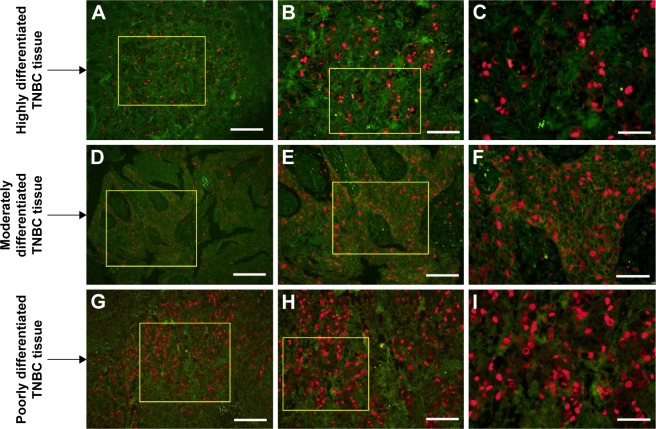
Immunofluorescent imaging of TOP2A.
Notes: TOP2A (red immunofluorescent signal by QD-605) in the cell nucleus was visible at high resolution against the clear discernible background. (A–C) The expression of TOP2A was less in highly differentiated TNBC tissues. (D–F) The signal in the moderately differentiated TNBC tissues was strong and the distribution was dense. (G–I) The signal in the poorly differentiated TNBC tissues was strongest, and the distribution was densest. The yellow box was used to show the magnified area for the next image. Scale bar =100 μm (A, D, and G), 50 μm (B, E, and H), and 25 μm (C, F, and I).
Abbreviations: TOP2A, topoisomerase 2 alpha; TNBC, triple-negative breast cancer.

