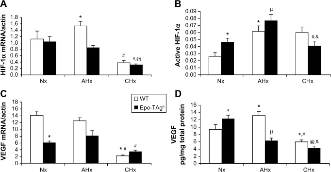
Figure 1.
Cardiac angiogenesis in Epo-TAgh mice.
Notes: HIF-1α and VEGF mRNA (A, C) and protein (B, D) expression in the heart of WT (white bar) and Epo-TAgh (black bar) mice in normoxia and after 48 hours (AHx) or 14 days (CHx) of hypoxic exposure. Epo deficiency led to a rise of angiogenesis through the HIF-1α and VEGF pathway activation. Epo-TAgh mice were not able to maintain cardiac adaptation to hypoxia during the long-term exposure. Values are expressed as mean ± SEM. *P<0.05 vs Nx WT; &P<0.05 vs Nx Epo-TAgh; #P<0.05 CHx vs AHx; @P<0.05 CHx Epo-TAgh vs CHx WT. μP<0.05 AHx Epo-TAgh vs AHx WT. Reprinted from Respir Physiol Neurobiol, Volume 186(2), El Hasnaoui-Saadani R, Marchant D, Pichon A, et al, Epo deficiency alters cardiac adaptation to chronic hypoxia, pages 146–154. Copyright 2013 with permission from Elsevier.20
Abbreviations: AHx, acute hypoxia; CHx, chronic hypoxia; Epo, erythropoietin; Epo-TAgh mice, Epo-deficient mice; HIF-1α, hypoxia-inducible factor-1α; Nx, normoxia; SEM, standard error of the mean; VEGF, vascular endothelial growth factor; WT, wild type.

