Figure 1.
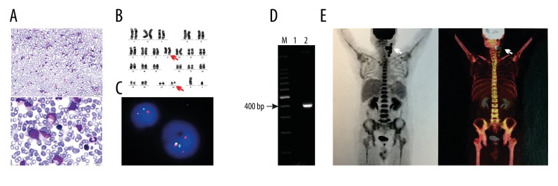
Morphology, cytogenetics, and molecular features of bone marrow and 18F-FDG PET/CT imaging analysis. (A) Bone marrow aspiration revealing a hypercellular marrow with the presence of 6.4% immature lymphocytes. (B) A G-banded karyotype of bone marrow showing the Ph chromosome: 46 XY, t(9;22)(q34;q11) (arrows). (C) FISH on the interphase nuclei of bone marrow using the Vysis Extra Signal probe revealing the typical p210 breakpoint in CML yielding 1 green, 1 red, 1 residual red, and 1 red-green fusion signal. (D) RT-PCR analysis of the mRNA extracted from bone marrow demonstrating p210 BCR/ABL1 rearranged bands (397 bp). Lane 1, marker; Lane 2, distilled water; Lane 3, bone marrow. (E) 18F-FDG PET/CT scanning of the body showing abnormal FDG accumulation in the left side of the neck and submandibular lymph nodes (arrows).

