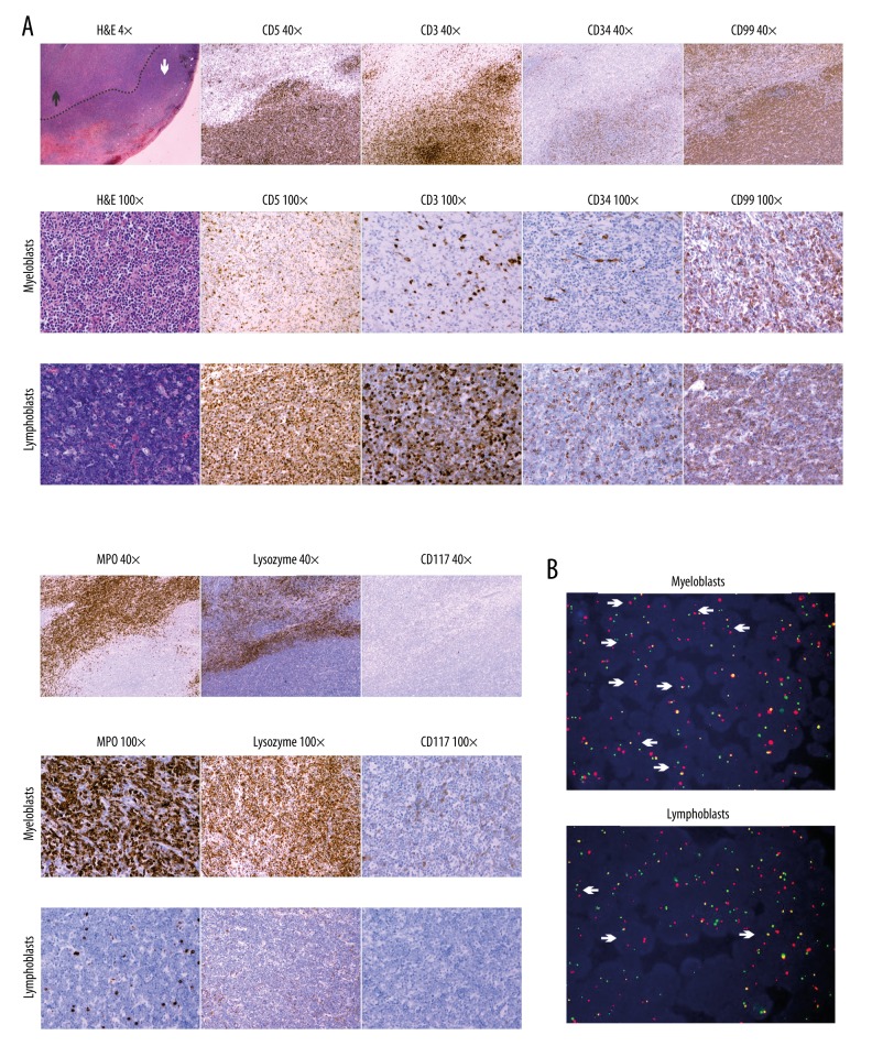
Figure 2.
Histopathology features of the lymph nodes. (A) Microscopic appearance showing the normal architecture of the lymph node substituted by diffuse infiltration of 2 populations of blastic cells with a clear dividing line – the red and sparse staining myeloblasts region (white arrow) and the blue and dense staining T lymphoblasts region (black arrow) (H&E, 4×). The sparse staining region is composed of neoplastic cells with myeloid differentiation, with background rich in blood vessels (H&E, 100×). The cells are strongly positive for MPO and lysozyme, and negative for CD117, CD5, CD3, CD34, and CD99 (immunohistochemistry with hematoxylin counterstain, 40× and 100×). The dense staining region is composed of T lymphoblasts with dense nuclear chromatin and a high nuclear-to-cytoplasmic ratio, and scattered macrophages with starry-sky appearance (H&E, 100×). The cells are strongly positive for CD5, CD3, and CD99; focally positive for CD34; and negative for MPO, lysozyme, and CD117 (immunohistochemistry with hematoxylin counterstain, 40× and 100×). (B) FISH on the paraffin-embedded biopsy specimen using the Vysis Extra Signal probe revealing the presence of p210 breakpoint in both myeloblasts (low cell density area) and T lymphoblasts (high cell density area). Positive cells (arrows) contain 1 green, 1 red, 1 residual red, and 1 red-green fusion signal.

