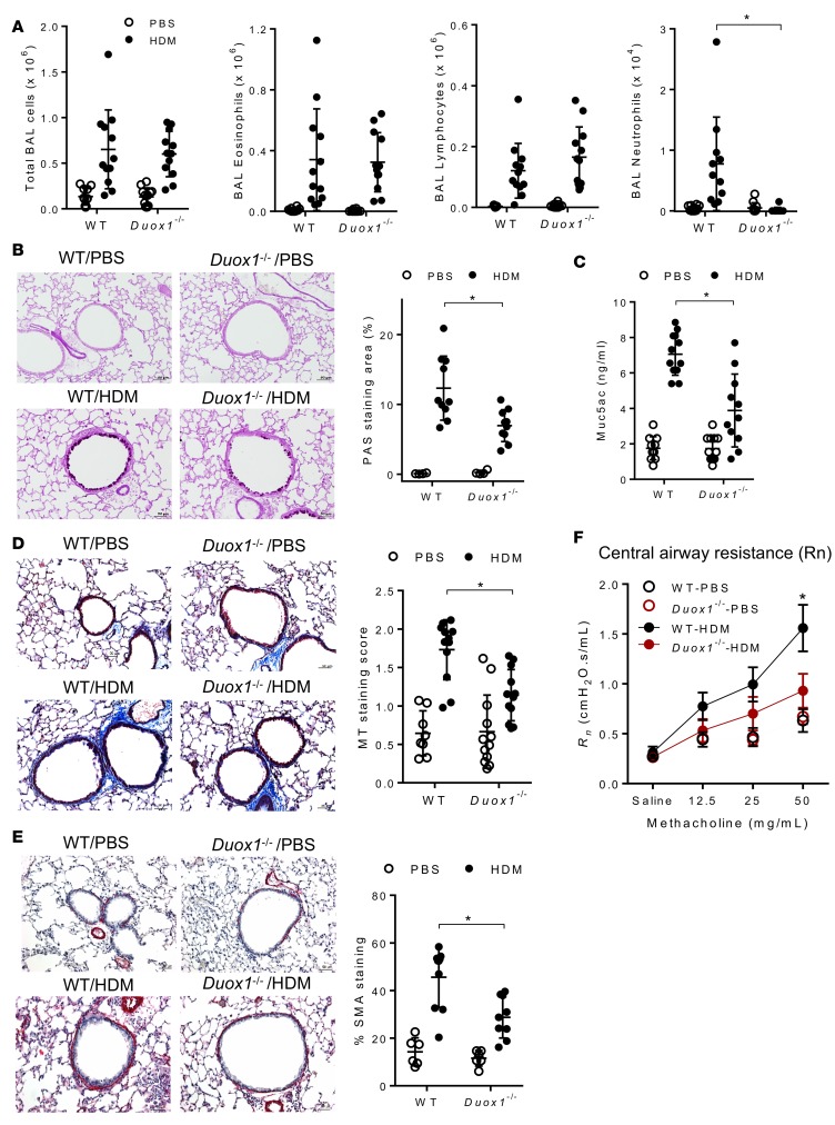
Figure 3. DUOX1 contributes to house dust mite–induced neutrophilic inflammation, mucous metaplasia, subepithelial fibrosis, and airways hyperresponsiveness.
(A) Quantification of total and differential cell counts in BAL fluids. (B) Analysis of mucous metaplasia by histochemical analysis of PAS staining and quantification of positive staining areas using Metamorph software. (C) Quantification of Muc5ac protein in BAL fluids by ELISA. (D and E) Evaluation of subepithelial fibrosis by Masson’s trichrome staining (D) or α-SMA immunostaining (E), as quantified by staining score or percentage of staining around airways. Scale bars: 50 μm. (F) Analysis of airways central airway resistance (Rn) in response to methacholine challenge. Representative histochemical images are shown, and dot plots represent mean ± SD of 8–12 replicates from 3 independent experiments. *P < 0.05 compared with corresponding controls, using 2-way ANOVA.

