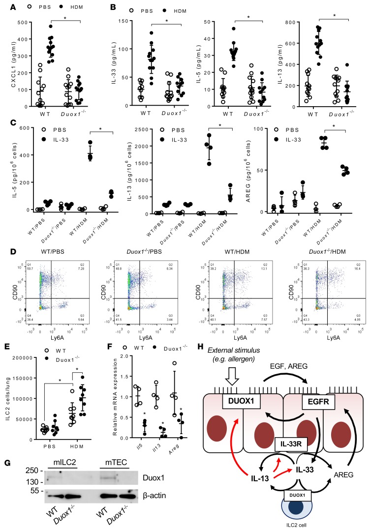
Figure 4. DUOX1 mediates type 2 cytokine responses and ILC2 activation during house dust mite–induced inflammation.
(A and B) Analysis of BAL cytokine levels of CXCL1 (KC) (A) and of type 2 cytokines, IL-33, IL-5, and IL-13 (B). (C) Analysis of IL-33–induced (10 ng/ml) production of ILC2-specific cytokines (IL-5, IL-13) and AREG from lung single-cell suspensions of PBS- or HDM-treated mice. (D and E) Flow cytometry analysis of ILC2 populations in lung tissues. (F) RT-PCR analysis of flow-sorted lung ILC2 populations from IL-33–triggered mice for type 2 cytokines, Areg, and Duox1. (G) Representative western blot analysis of Duox1 protein and β-actin in sorted ILC2 cells (mILC2) and primary mTECs. (H) Schematic representation of proposed interactions among DUOX1, EGFR, IL-33, and IL-13 within the airway. Black arrows reflect enzyme activation or stimulation of cytokine secretion, and red arrows indicate effects on mRNA expression. Dot plots represent mean ± SD of 8–12 replicates from 2–3 separate experiments. *P < 0.05 compared with corresponding controls by 2-way ANOVA.

