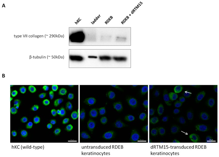
Figure 8.
Analysis of type VII collagen expression. (A) Western blot analysis revealed an increase in type VII collagen expression (~290 kDa) in dRTM15-transduced RDEB keratinocytes in comparison to untransduced RDEB keratinocytes showing less type VII collagen expression. Human wild-type keratinocytes showed high type VII collagen levels and served as a control (hKC). As a protein loading control β-tubulin was used. For dRTM treated and untreated RDEB keratinocytes similar protein amounts were loaded onto the gel; (B) Immunofluorescense staining of wild-type keratinocytes, untransduced and RTM15-transduced patient keratinocytes. Arrows indicate dRTM15-transduced RDEB keratinocytes showing higher amounts of type VII collagen expression (green) compared to untransduced patient cells. Cell nuclei were 4′,6-diamidin-2-phenylindol (DAPI) stained (blue). hKC: human wild-type keratinocytes; RDEB: patient keratinocytes harbouring the homozygous mutation within exon 80. Scale bars represent 20 μm.

