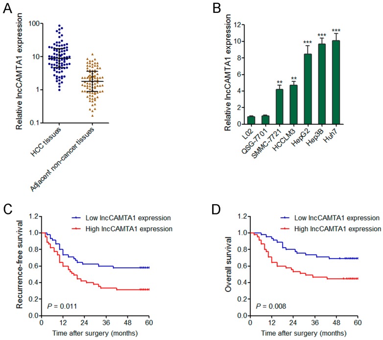
Figure 3.
lncCAMTA1 is increased in HCC and indicates poor outcome. (A) lncCAMTA1 is increased in HCC tissues in comparison with paired adjacent non-cancerous hepatic tissues. Data are shown as median with interquartile range. n = 90, p < 0.001 by Wilcoxon signed-rank test; (B) lncCAMTA1 is increased in HCC cell lines SMMC-7721, MHCC97H, HCCLM3 and HepG2, in comparison with normal liver cell lines L02 and QSG-7701. Data are shown as mean ± SD from at least three independent experiments. ** p < 0.01, *** p < 0.001 by Student’s t test; (C,D) Kaplan–Meier analyses indicated that patients with high expression of lncCAMTA1 had a shorter recurrence-free survival (p = 0.011, log-rank test); (C) and overall survival (p = 0.008, log-rank test); (D) time than those with low expression of lncCAMTA1.

