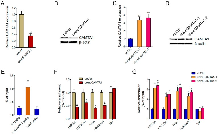
Figure 6.
lncCAMTA1 inhibits CAMTA1 expression via changing chromatin structure at the CAMTA1 promoter. (A) CAMTA1 mRNA levels in HCCLM3 cells stably overexpressing lncCAMTA1 and control HCCLM3 cells; (B) CAMTA1 protein levels in HCCLM3 cells stably overexpressing lncCAMTA1 and control HCCLM3 cells; (C) CAMTA1 mRNA levels in lncCAMTA1 stably depleted and control HepG2 cells; (D) CAMTA1 protein levels in lncCAMTA1 stably depleted and control HepG2 cells; (E) Chromatin isolation by RNA purification (ChIRP) assays showed that lncCAMTA1 has a significantly genomic occupancy on the CAMTA1 promoter; (F,G) Chromatin immunoprecipitation (ChIP) assays were performed using H3K9ac, H3K27ac, H4ac, H3K4me3, or non-specific IgG antibodies in HCCLM3 cells stably overexpressing lncCAMTA1 or control HCCLM3 cells (F), lncCAMTA1 stably depleted or control HepG2 cells (G). The retrieved DNA was amplified by quantitative PCR (qPCR) to measure the occupancy of indicated histone marks at the CAMTA1 promoter. For all panels, data are shown as mean ± SD. * p < 0.05, ** p < 0.01 by Student’s t test.

