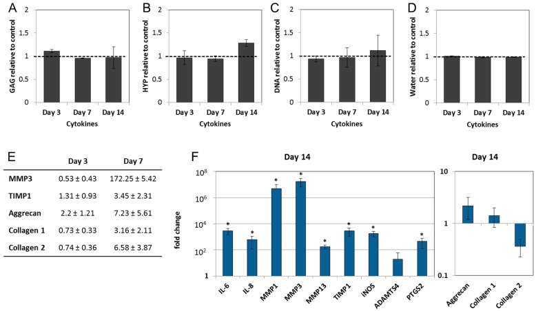
Figure 4.
IL-1β and TNF-α induce an inflammatory and catabolic phenotype in the NPs: (A) Glycosaminoglycan; (B) collagen; (C) DNA; and (D) water content in cytokine-treated NPs remained unchanged over a period of 14 days, when compared with the untreated group (set as 1). Graphs show ECM composition relative to untreated samples (mean ± SEM from n = 4); (E) inflammatory and catabolic gene expression was not detectable on Days 3 and 7, and the expression of MMP3, TIMP1 and ECM macromolecules were close to the control values (fold change); (F) on Day 14, IL-1β and TNF-α significantly induced gene expression of interleukins (IL-6, IL-8), collagenases (MMP1, MMP3, MMP13) and their tissue inhibitor (TIMP1), aggrecanase 1 (ADAMTS4) and pain mediators (PTGS2, iNOS). Graphs show fold change relative to the untreated samples (mean ± SEM from n = 4–7). * p < 0.05 vs. untreated control (ANOVA).

