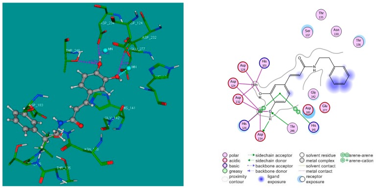
Figure 5.
3D (left); and 2D (right) binding modes of CAPA (1) inside the active site of h-ARG I. For the 3D binding mode, CAPA is represented as a “ball and stick” form: C (grey), O (red), and H (white); manganese ions are represented as cyan spheres; and the crucial residues of the binding site are represented by lines. H-bonds are represented as pink dotted lines, while the metal coordination bonds are represented by cyan dotted lines. Docking simulations were performed by FlexX program implemented in LeadIt 2.0.2 software, and the pictures of docking solution were created by MOE 2008.10 software.

