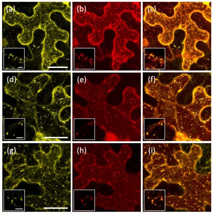
Figure 7.
Co-expression of AtRMR2-RFP with either AtRMR1-YFP or its deletion mutants. Stacked confocal images, excepted inserts (a–i; single images), of epidermal cells expressing the following combinations: (a–c) AtRMR1-YFP and AtRMR2-RFP; (d–f) AtRMR1ΔRing-YFP and AtRMR2-RFP; and (g–i) YFP-AtRMR1ΔPA and AtRMR2-RFP. YFP signal (a,d,g); RFP signal (b,e,h); and merged images (c,f,i). Scale bars = 30 µm; and 5 μm (inserts).

