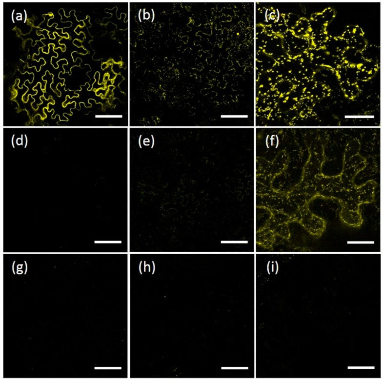
Figure 8.
Dimerization of AtRMRs tested by BiFC. Confocal images of epidermal cells expressing split-YFP constructs: (a) The positive control, p6-nYFP and p6-cYFP; (b,c) AtRMR2ΔRingSer-nYFP and AtRMR2ΔRingSer-cYFP; (d) AtRMR1ΔRing-nYFP and AtRMR1ΔRing-cYFP; (e–f) AtRMR2ΔRingSer-nYFP and AtRMR1ΔRing-cYFP; (g) negative control, AtRMR2ΔRingSer-nYFP alone; (h) negative control, AtRMR2ΔRingSer-cYFP alone; and (i) negative control, AtRMR2ΔRingSer-nYFP and p6-cYFP. Singles images showing an overview of leaf tissue expressing the indicated fusion proteins (a,b,d,e,g–i), excepted stacked confocal images showing the localization in punctate structures of AtRMR2ΔRingSer homodimers and AtRMR2ΔRingSer-AtRMR1ΔRing heterodimers in a single epidermal cell, respectively (c,f). YFP signal (a–i). Scale bars = 100 μm (a,b,d,e,g–i); 30 µm (c,f).

