Figure 5.
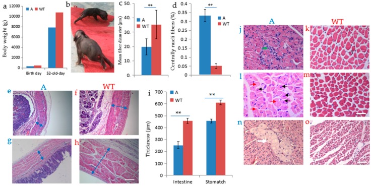
DMD deficiency resulted in pathological alterations in the DMD-modified pig. (a) The birth weight (480 g) and body weight at 52 days (7809 g) of founder A were lower than that of the wild-type piglet (birth weight 575 g and body weight at 52 days was 110,200 g); (b) Behavioral performance of muscle weakness of founder A; (c) Mean minimal Feret’s diameter of muscle fibers in biceps femoris muscle of 52-day-old founder A and WT; (d) Proportion of muscle fibers with central nuclei in deltoid muscle of 52-day-old founder A and WT. ** p < 0.01; (e–h,j–o) Haematoxylin and eosin (H&E) staining, paraffin section; (e–i) Smooth muscle thickness of stomach (e) and intestine (g) were dramatically decreased compared with that of WT (f,h). Blue arrows indicate the thickness of stomach and intestine; (j) Cross-section of biceps femoris muscle revealed disordered myofiber construction and the green arrow indicated the aggregated nuclei; (l) Black arrows indicated the rounded myofibers with central nuclei, and red arrows indicated the necrosis of myofibers in deltoid muscle; (n) Multifocal area of pale discoloration in cardiac muscle was observed in section; (k,m,o) histological section of skeletal muscle and cardiac muscle of wild-type. Scale bars: 50 µm (e–h,j–m),100 µm (n,o).

