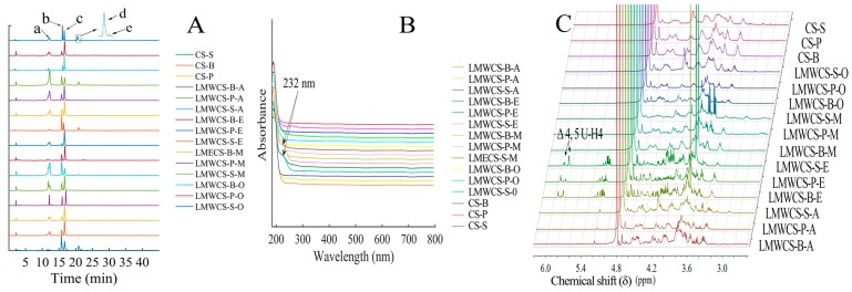
Figure 1.
Characterization of low molecular weight chondroitin sulfate (LMWCS) samples. (A) Disaccharide compositions of all CS samples from three sources depolymerized by different methods. B, P and S stand for bovine, porcine and shark cartilage, respectively. A stands for the HCl method; E stands for enzymatic method; M stands for microwave-assisted alkaline method; and O stands for oxidative method. a, ΔDi-0S; b, ΔDi-6S; c, ΔDi-4S; d, ΔDi-2,6diS; e, ΔDi-4,6diS; (B) UV spectra of all CS samples; (C) Proton nuclear magnetic resonance (1H-NMR) spectra of chondroitin sulfates (CSs) from three sources and low molecular weight chondroitin sulfates (LMWCSs) depolymerized by different methods.

