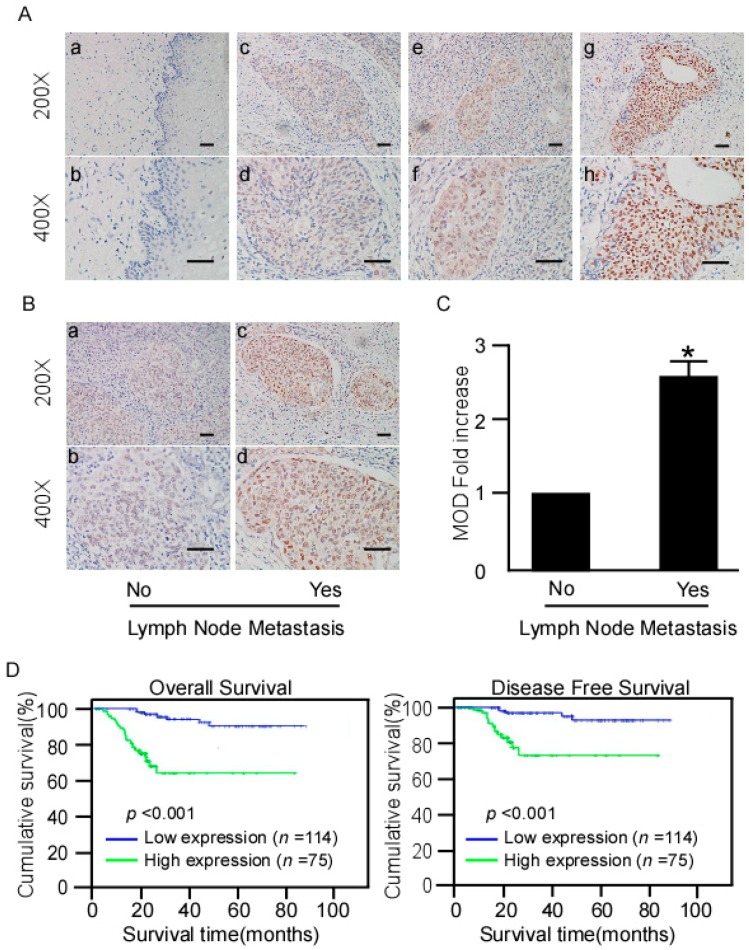
Figure 3.
IHC detection of NR2F6 protein expression in paraffin-embedded cervical cancer tissues. Positive NR2F6 staining was observed mainly in the nuclei. (A) (a,b) NR2F6 expression was not detected in normal cervical tissues; (c,d) representative images of weak NR2F6 staining in cervical cancer tissues; (e,f) representative images of moderate NR2F6 staining in cervical cancer tissues; (g,h) representative images of strong NR2F6 staining in cervical cancer tissues(scale bars = 50 μm); (B,C) Statistical analyses of the average MOD (mean optical density) of NR2F6 staining in the LNM (Lymph node metastasis) and LNM-free groups. Scale bars = 50 μm, * p < 0.05; (D) Kaplan–Meier curves of univariate analysis data (log-rank test) showing the OS (overall survival) and DFS (disease-free survival) for patients with high versus low NR2F6 expression.

