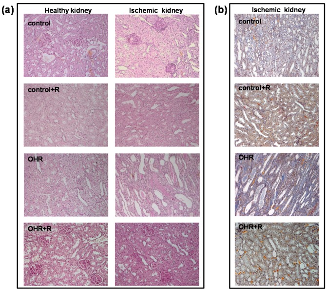
Figure 5.
Representative PAS (a) and trichrome (b) staining images (magnification 200×) of renal sections from control and OHR rats. The histological analysis indicated the appearance of morphological alterations, including mesangial hypercellularity, mesangial matrix expansion, tubular casts, tubular dilation, interstitial infiltration, and interstitial fibrosis. These alterations were more evident in ischemic kidneys of OHR rats compared to controls and were reverted by rostafuroxin treatment. The evaluation of histological parameters was scored semi-quantitatively as shown in Table 3. The glomerular evaluation was performed on 30 glomeruli per section, indicating that it was modestly affected by ischemia and rostafuroxin (score in Table 3).

