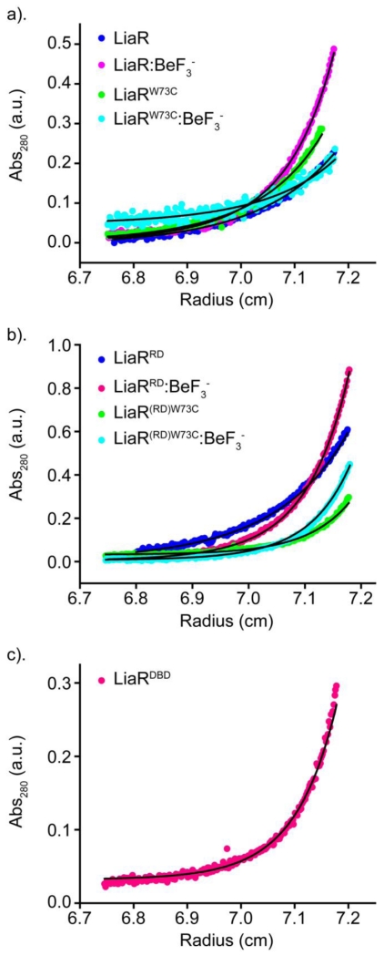
Figure 1. Beryllofluoride (BeF3−) promotes formation of Efm LiaR dimers.
Sedimentation equilibrium analytical ultracentrifugation (AUC) profiles for: (a) full length Efm LiaR (dark blue); Efm LiaR with BeF3− (magenta); adaptive mutant Efm LiaRW73C (green); and adaptive mutant Efm LiaRW73C with BeF3− (cyan). (b) AUC profiles for Efm LiaR(RD) (blue); Efm LiaR(RD) with BeF3− (magenta); Efm LiaR(RD)W73C (green); and Efm LiaR(RD)W73C with BeF3− (cyan): (c) AUC profile for the DNA binding domain of Efm LiaR (LiaR(DBD)) alone (magenta). For each protein, the SEQ profiles were globally fitted to a monomer only or monomer-dimer equilibrium model.

