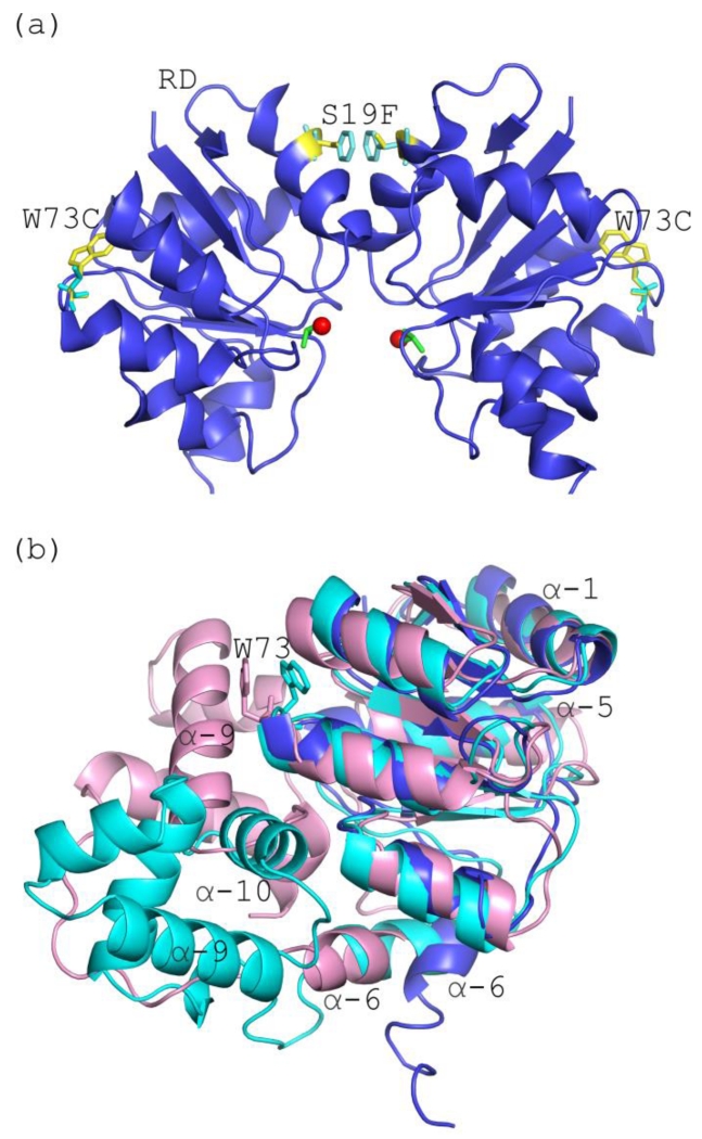
Figure 7. Mutations associated with daptomycin resistance are located within the receiver domain.
(a) Expanded view of the Efm LiaR receiver domains showing the positions of the mutations W73C and S19F. The S19F mutation (yellow/cyan sticks) is within the dimerization interface, while the adaptive mutation W73C (yellow/cyan sticks) is not located within the molecular surfaces that comprise the LiaR dimer: (b) Structural alignment of the activated Efm LiaR receiver (blue) compared to the inactive state structures of VraR/NarL receiver domains (cyan/light pink). Position 73 in the VraR and NarL structures are depicted as sticks (cyan/light pink, respectively).

