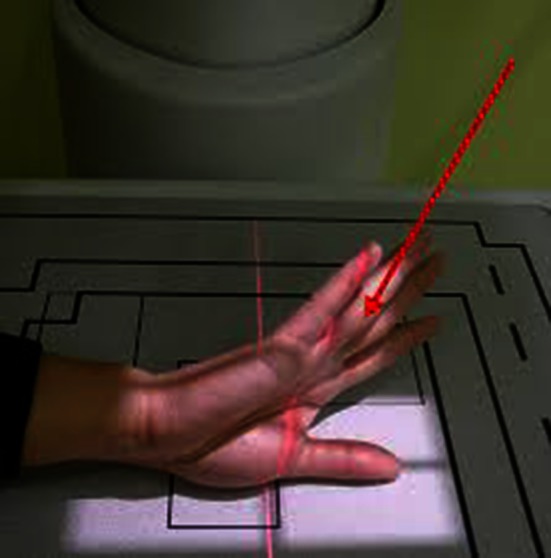
Fig. 2.

The Robert’s view radiograph [16] is taken with the wrist in maximal pronation, with the dorsum of the thumb parallel to the table, and the imaging beam is centered on the trapeziometacarpal joint (Published with permission from Amy L. Ladd MD.).
