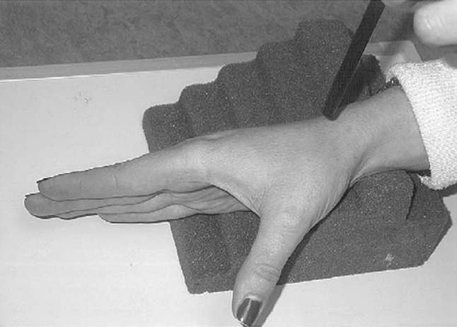
Fig. 3.

The Bett’s view (Gedda’s view) radiograph is taken with the hand pronated 30° and the imaging beam directed obliquely in a distal to proximal direction and centered over the trapeziometacarpal joint. (Published with permission from Dela Rosa TL, Vance MC, Stern PJ. Radiographic optimization of the Eaton classification. J Hand Surg Br. 2004;29:73–177.).
