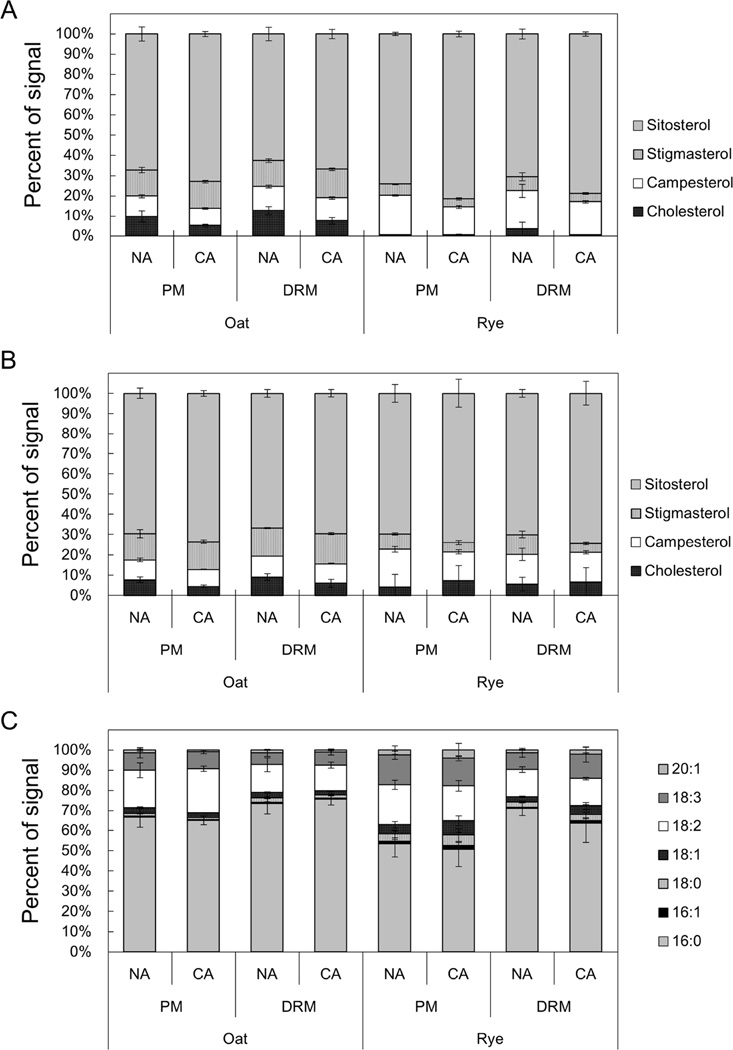
Fig. 4. Sterol and fatty acyl compositions of SG and ASG in PM and DRM fractions.
SG and ASG in PM and DRM were quantified by direct infusion MS/MS analysis. The Y-axis of the graph represents the proportion of signals derived from each sterol or acyl species in the total SG or ASG signals. Error bars indicate standard deviations (n=4). Molecular species compositions of the sterol part of SG and ASG are shown in A and B, respectively. C shows molecular species compositions of acyl chains of ASG.

