Fig. 1.
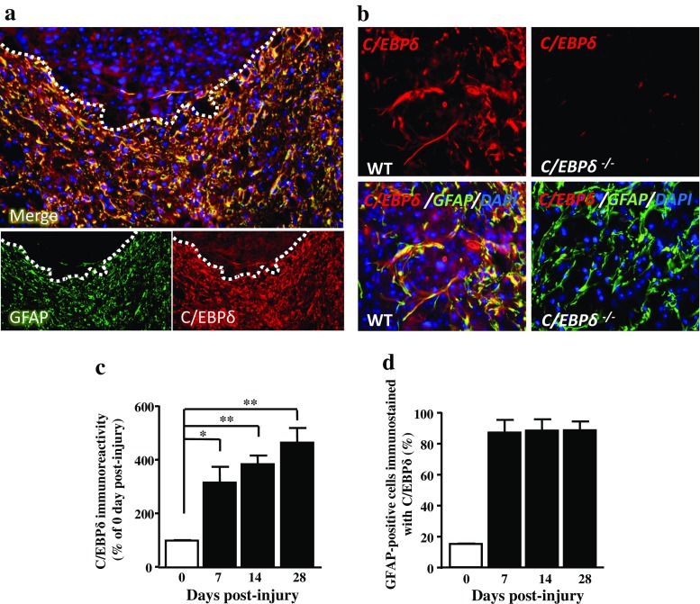
C/EBPδ is associated with GFAP-positive astrocytes in the glial scar of wild-type mice after SCI. a Transverse sections of the spinal cord obtained from wild-type mice are immunostained with anti-GFAP and -C/EBPδ antibodies at 14 days after spinal cord injury. Both GFAP and C/EBPδ immunoreactivity is apparent in the residual cord tissue (dotted lines) ventrolateral to the lesion epicenter particularly along the lesion border. b At higher magnification, co-localization of GFAP and C/EBPδ immunoreactivity is evident in astrocytes of wild-type mice, whereas C/EBPδ immunoreactivity is negative in astrocytes of C/EBPδ−/− mice. c Quantitative analyses (n = 6 for each time point) show that the intensity of C/EBPδ immunoreactivity increases significantly over time in the injured spinal cord. d The percentage of GFAP-positive astrocytes that is co-localized with C/EBPδ immunoreactivity increases from 7 days post-injury onwards

