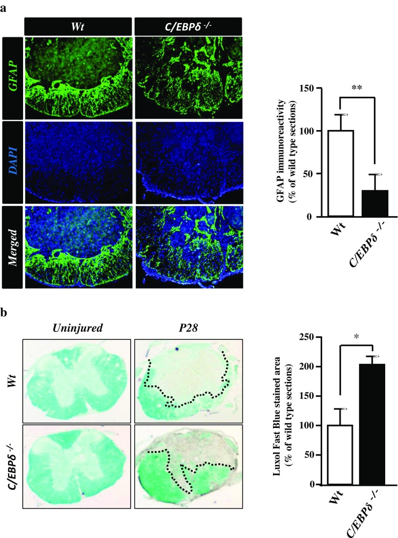
Fig. 3.
C/EBPδ −/− mice display less severe glial scar formation and more residual white matter than wild-type mice after SCI. a Transverse sections of the spinal cord are immunostained with anti-GFAP antibody 28 days after SCI. GFAP-positive astrocytes of the C/EBPδ −/− mice are relatively dispersed, loosely aggregated in the glial limitans where the glial scar is formed compared with those of the wild-type mice, suggesting a fragmental, less severe glial scar in the C/EBPδ −/− mice. Statistical analysis reveals that the intensity of GFAP immunoreactivity is lower in the C/EBPδ −/− mice than in the wild-type mice (n = 6 per genotype). b Transverse sections of the spinal cord are stained with Luxol Fast Blue to visualize the residual white matter around the lesion epicenter 28 days after SCI. C/EBPδ −/− mice show more prominent Luxol Fast Blue staining and quantitatively larger area of residual white matter than the wild-type mice (n = 6 per genotype). Dotted lines demarcate the residual cord tissue

