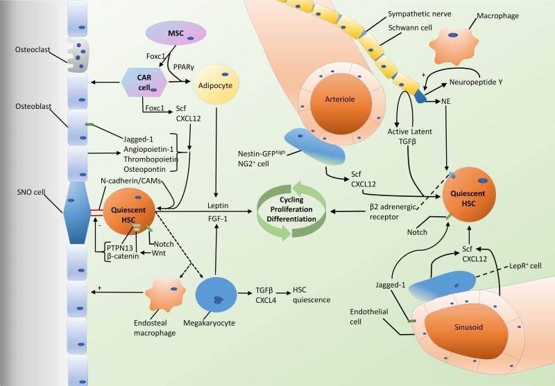
Figure 1. The BM HSC microenvironment.
In the osteoblastic niche near the bone surface several stromal cells as well as HSC-derived cells regulate HSC function. Scf, CXCL12, and several other secreted factors from stromal cells, in addition to direct interactions via CAMs promote HSC quiescence. Wnt effectors PTPN13 and β-catenin and signals from adipocytes and megakaryotes including leptin and FGF-1, respectively, may promote HSC differentiation and expansion. In the vascular niche there is controversy over the roles of arteriolar and sinusoidal zones, with evidence supporting the association of both with the quiescent HSC niche. Endothelial cells, sympathetic nerves, Schwann cells, macrophages, LepR+ cells, and Nestinhigh NG2+ stromal cells all play a role in regulating HSC function via multiple mechanisms including Scf and CXCL12 secretion, catecholaminergic signaling, TGFβ, and the Notch pathway.

