Fig. 1.

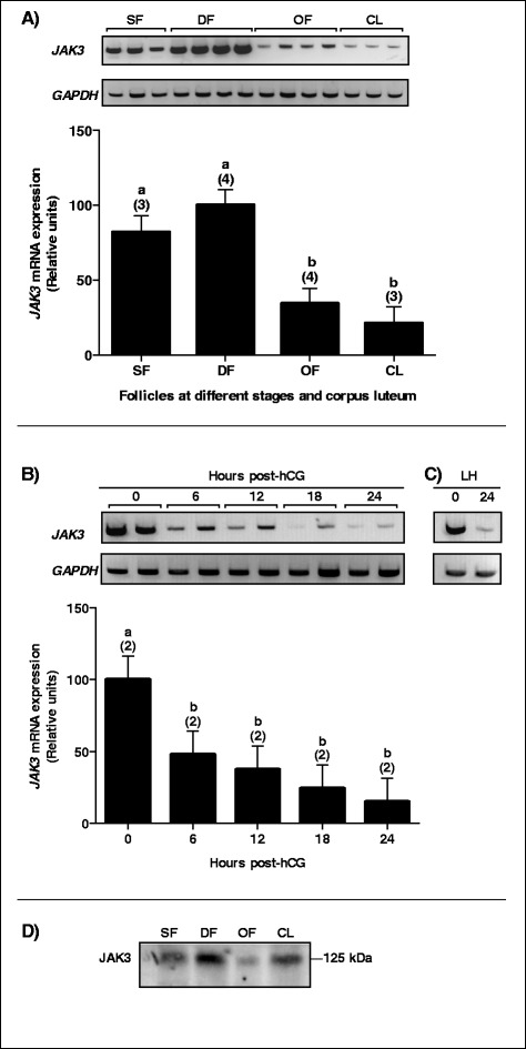
JAK3 mRNA expression and regulation in bovine follicles. Total RNA extracts of GC from small follicles (SF), dominant follicles (DF), ovulatory follicles (OF), and corpus luteum at day 5 (CL) were analyzed by RT-PCR for JAK3 with GAPDH used as reference. a Gel analysis of JAK3 in different groups of follicles and CL and corresponding histograms. JAK3 mRNA expression was strongest in DF and was significantly decreased in OF and CL (P < 0.0001). b Gel analysis of JAK3 mRNA regulation in hCG-induced follicular walls (FW) isolated from OF at 0, 6, 12, 18 and 24 h (hrs) after hCG injection and corresponding histograms. JAK3 mRNA was markedly decreased in FW 6 h post-hCG compared to its expression before hCG treatment (P < 0.01) and reached its weakest expression 24 h post-hCG as compared to 0 h (P < 0.0001). c Gel analysis of JAK3 mRNA regulation by endogenous LH. Similar to hCG, a decrease in JAK3 expression was observed 24 h after endogenous LH (LH) surge as compared to 0 h. d Representative JAK3 protein expression and regulation in bovine follicles. Total protein extracts of GC from SF, DF, OF, and CL were analyzed by western blot using anti-JAK3 antibodies. The strongest JAK3 protein expression was observed in the DF while weakest expression was observed in OF reflecting the regulation of the mRNA. Data are presented as least-square means ± SEM, and the number of independent samples per group is indicated in parenthesis
