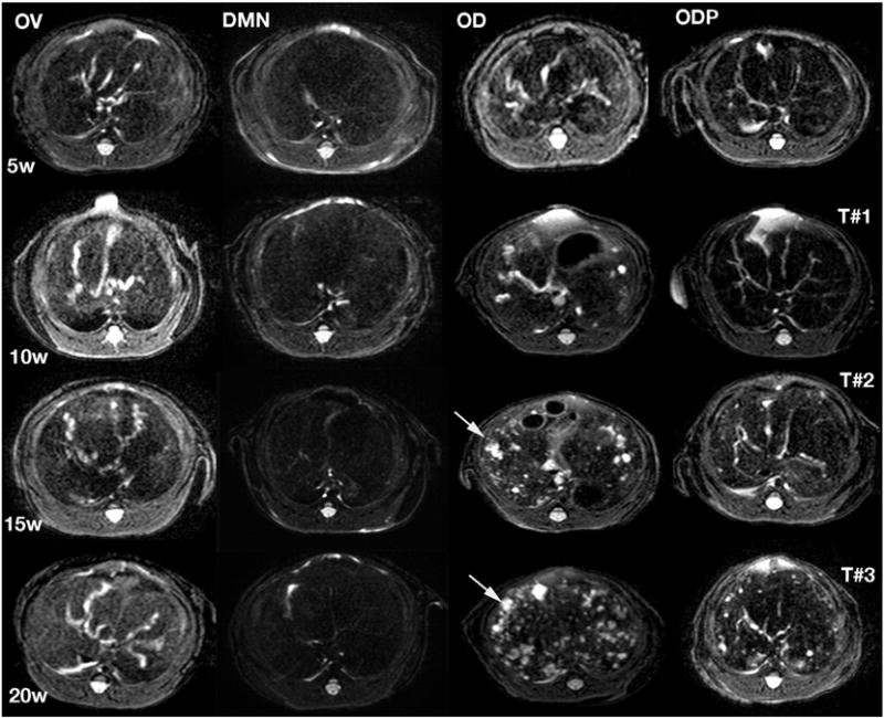
Fig. 2.

Axial T2 weighed images of hamsters in the OV, DMN, OD and ODP groups. Experimental weeks (W) and order of MRI examination (T#) are labeled on the left and right sides of the figure, respectively. The first MRI (T#1) applied at week five (5W) being shown in the first row could reveal different degrees of hepatobiliary lesions. The animals in the OD group showed the most progressive appearance. Arrows indicated periductal fibrosis and duct dilatation, which increased in the OD hamsters. Hamsters of the ODP group exhibited less severe lesions compared to hamsters in the DMN and OV groups. Progression of the hepatobiliary lesions is in accordance with number of cycles of infections with O. viverrini and treatment with PZQ treatment: the higher number, the more progress.
