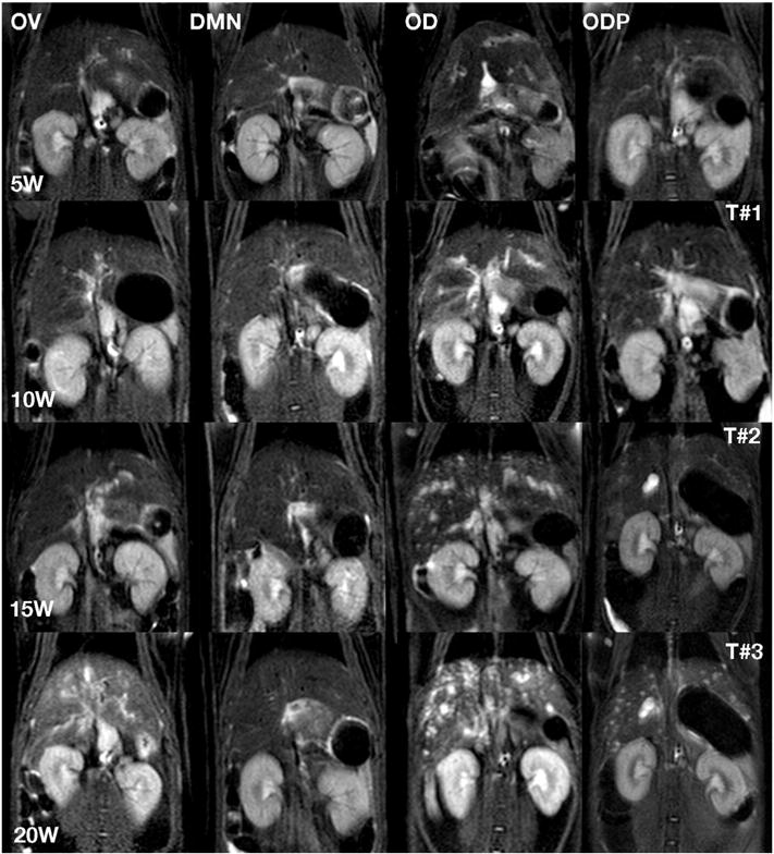
Fig. 3.

Three coronal planes of MRI scans (T#1, T#2, T#3) at weeks 5, 10, 15 and 20 (5W, 10W, 15W and 20W) after infection for the treatment groups, OV, DMN, OD and ODP. These T2 weighed images documented pathological changes similar to those revealed in Fig. 1. The lesional changes in the ODP livers in the final MRI scan (T#3) were not as markedly progressive as those in the OD liver.
