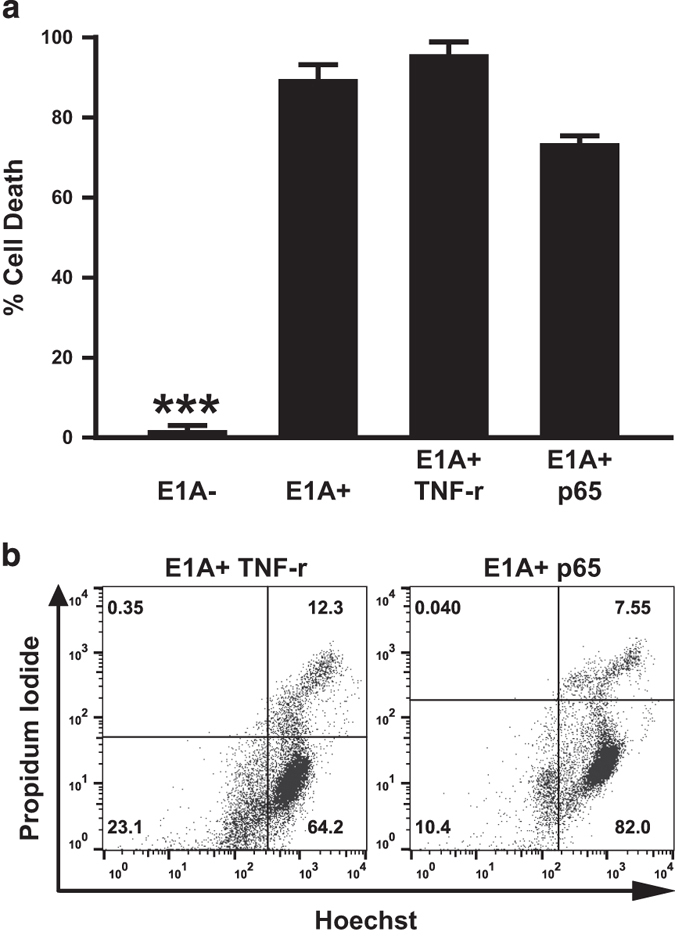
Figure 2.

Role of NF-κB cellular responses in etoposide-induced apoptosis. (a) Four different mouse cell lines – E1A− controls; E1A+ controls; TNF-resistant E1A+ cells (E1A+ TNFr); or p65/RelA-overexpressing E1A+ cells (E1A+ p65) cells – were treated with 400 μM etoposide for 18 h. Cell viability was determined by MTS staining and expressed as % cell death (mean±S.E.M.; n=3, ***P<0.001). (b) Hoechst/PI staining patterns of E1A+ TNFr or E1A+ p65 cells following treatment with etoposide (400 μM) for 18 h.
