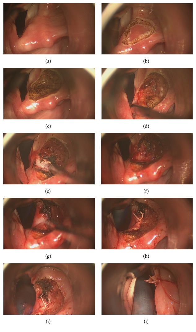
Figure 2.
(a) Right arytenoid cartilage is visualized with modified laryngoscope; intubation tube is elevated with this laryngoscope out of surgical field, thus enlarging field of vision. (b) Anteriorly based triangular incision was marked with CO2 laser spots on right arytenoid. (c) Mucosa covering arytenoid was removed revealing superior surface of cartilage. (d) Anterior half of arytenoid was dissected; mucosa medial to arytenoid was preserved. (e) Anterior half of arytenoid was cut with CO2 laser transversely and is about to be removed. (f) Anterior half of arytenoid was removed. Mucosa medial to arytenoid was preserved. (g) Posteromedially based advancement flap was outlined and shown. (h) Posteromedially based advancement flap was sutured posterolaterally. (i) After posteromedially based advancement flap is sutured posterolaterally, membranous vocal fold is about to be sutured posterolaterally. (j) Membranous vocal fold was sutured posterolaterally; glottis is enlarged.

