FIG 2.
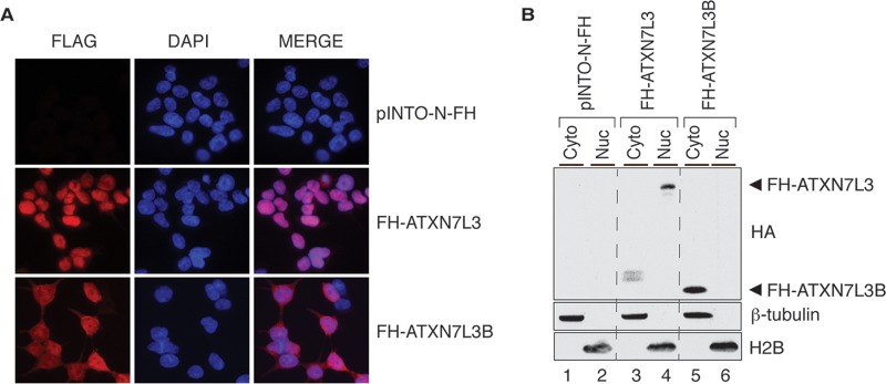
ATXN7L3B mainly localizes to the cytoplasm, whereas ATXN7L3 is nuclear. (A) Immunofluorescence using Flag antibody (red) on 293T cells stably expressing pINTO-N-FH vector, pINTO-N-FH-ATXN7L3, or pINTO-N-FH-ATXN7L3B. Nuclei were counterstained with DAPI (blue). (B) Cytoplasmic and nuclear fractions were isolated from 293T cells stably expressing pINTO-N-FH vector, pINTO-N-FH-ATXN7L3, or pINTO-N-FH-ATXN7L3B. Proteins were resolved by SDS-PAGE and detected by immunoblotting with the indicated antibodies. FH-ATXN7L3 and FH-ATXN7L3B proteins bands are indicated with arrowheads. H2B marks the nuclear fraction, while β-tubulin marks the cytoplasmic fraction.

