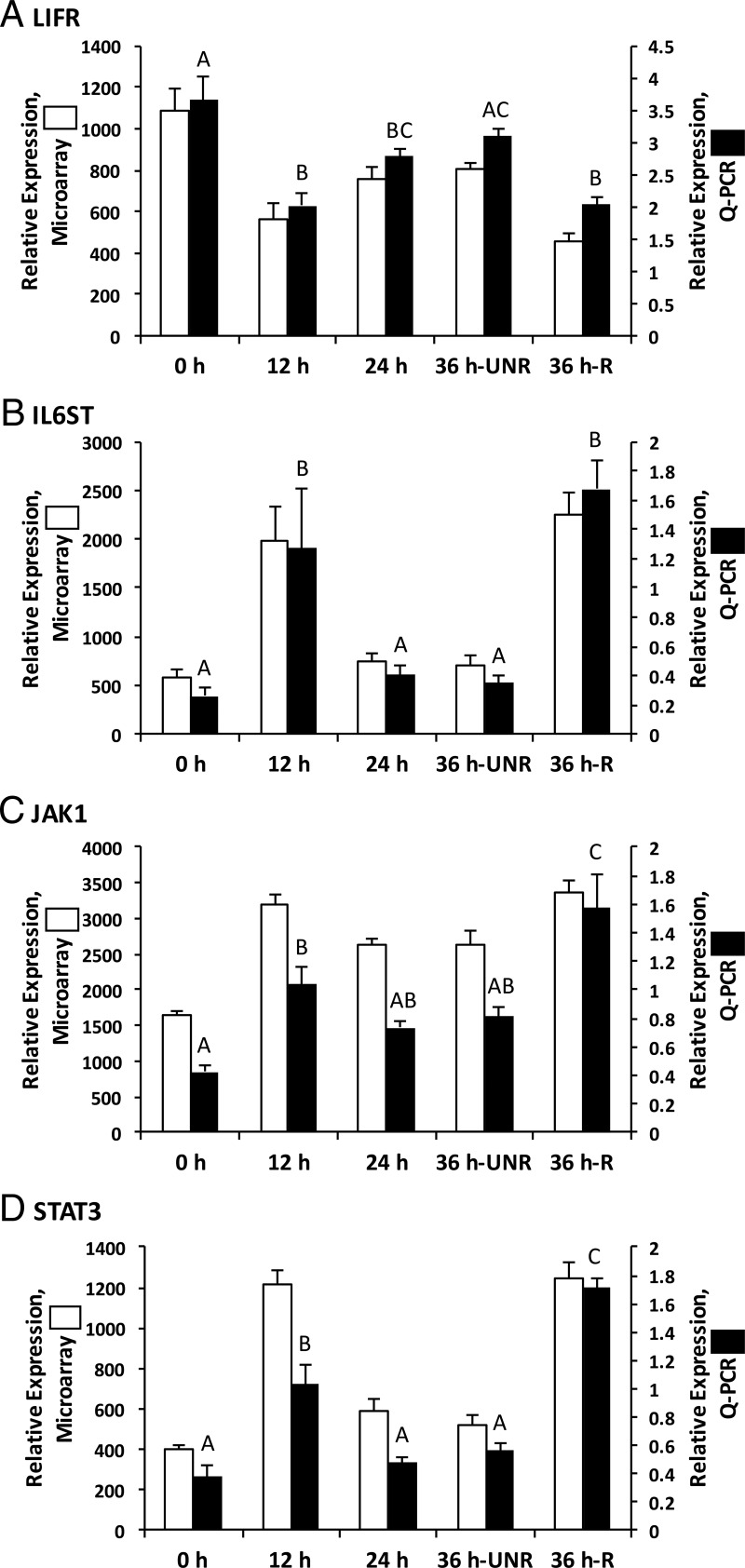
Figure 2.
The mRNAs corresponding to LIFR and its associated downstream signaling components are expressed in the rhesus macaque periovulatory follicle. Although LIFR mRNA levels show a moderate decline (1.8-fold) in the macaque follicle 12 hours after hCG administration (A), mRNAs encoding downstream LIF signaling components, including IL6ST (B), JAK1 (C), and STAT3 (D), exhibit a significant (P < .05) increase 12 hours after hCG administration relative to the pre-hCG time point. A secondary increase in the level of mRNAs encoding IL6ST, JAK1, and STAT3 was observed in ruptured rhesus macaque follicles obtained 36 hours after hCG administration. Columns with different letters are significantly different from one another (P < .05).

