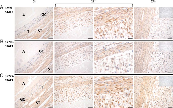
Figure 4.
Total and phosphorylated STAT3 levels increase in the rhesus macaque follicle after an ovulatory stimulus. Immunostaining intensity of total STAT3 (A), STAT3 phosphorylated at tyrosine 705 (pY705) (B), and STAT3 phosphorylated at serine 727 (pS727) (C) is increased in the rhesus macaque follicle 12 hours (bracket, left and right panels: ×40 and ×100 objectives, respectively) and 24 hours after the injection of an ovulatory bolus of hCG relative to that of follicles obtained before hCG administration (0 h). The insets located in the 24-hour panel include the negative controls (eg, irrelevant isotype-matched antibody) for each primary antibody used. The various ovarian cell types and compartments are indicated in the panels, including antrum (A), granulosa cells (GCs), theca cells (T), and stroma (ST). Images are representative of 2–3 ovaries obtained at each time point (0, 12, and 24 h after hCG) from animals undergoing COv protocols. Scale bar, 50 and 20 μm for ×40 and ×100 magnification, respectively.

