Figure 4.
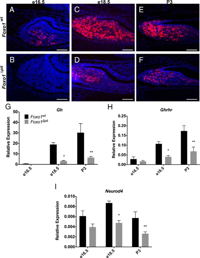
Terminal differentiation of somatotropes is delayed in Foxo1Δpit embryos. Immunohistochemistry for GH (red) was performed on coronal sections from Foxo1Δpit embryos and wild-type littermate controls at e16.5 (A and B), e18.5 (C and D), and P3 (E and F). All cell nuclei are stained with DAPI (blue). Images shown are representative of embryos from three different litters at e16.5, five different litters at e18.5, and four different litters at P3. Scale bars, 100 μm. RT-qPCR was performed for Gh (G), Ghrhr (H), and Neurod4 (I) using total RNA from Foxo1Δpit embryos and wild-type littermate controls at e16.5, e18.5, and P3. Values are relative to Actb for each sample. Data are expressed as mean ± SEM of five (Gh, Ghrhr) and four (Neurod4) animals of each genotype and each age. The data were analyzed by a two-way ANOVA with a Sidak's multiple comparisons test to determine significant differences between Foxo1Δpit embryos and wild-type littermate controls at each age. *, P < .05; **, P < .01.

