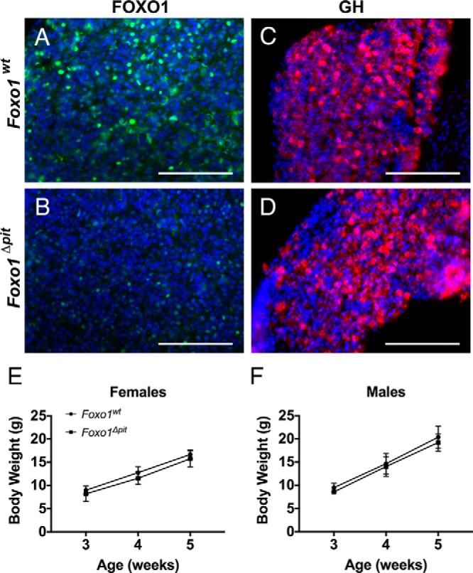
Figure 5.

Body weight is normal in Foxo1Δpit mice. Immunohistochemistry for FOXO1 (green)(A, B) or GH (red)(C, D) was performed on coronal sections from Foxo1Δpit mice and wild-type littermate controls at 3 weeks of age. All cell nuclei are stained with DAPI (blue). Images shown are representative of mice from at least three different litters. Scale bars, 100 μm. Foxo1Δpit mice and wild-type littermate control females (E) and males (F) were weighed at 3, 4, and 5 weeks of age. Data are expressed as mean ± SEM of three mice of each genotype. The data were analyzed by a two-way ANOVA with a Sidak's multiple comparisons test to determine significant differences between Foxo1Δpit mice and wild-type littermate controls.
