Figure 6.
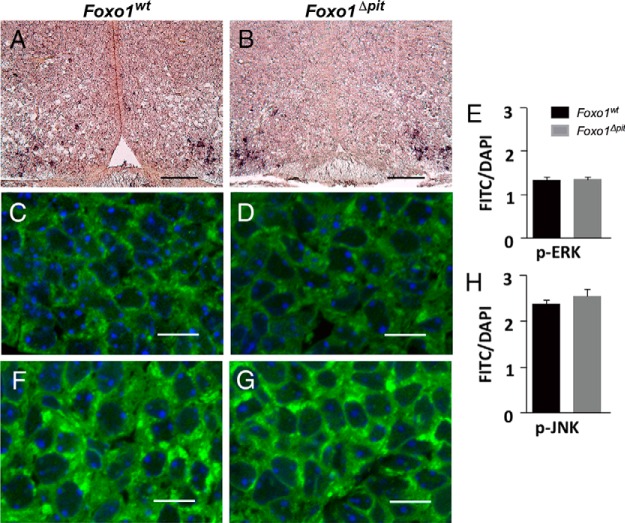
GHRH neurons are present in the arcuate nucleus of Foxo1Δpit embryos at e18.5. In situ hybridization was performed for Ghrh (dark brown) on coronal sections from wild-type embryos (A) and Foxo1Δpit littermates (B) at e18.5. Images shown are representative of embryos from two different litters for each genotype. Scale bars, 100 μm. To assess GHRH signaling, pituitary sections were immunostained for either p-ERK (C and D) or p-JNK (F and G) followed by the appropriate Alexa Fluor 488-conjugated secondary antibody (green) and then stained with DAPI (blue). Sections were imaged by confocal laser-scanning microscopy. Images are representative of sections from three different litters. Scale bars, 10 μm. A ratio of the fluorescence intensity of either p-ERK- (E) or p-JNK (H)-positive cells to total DAPI per pituitary section was calculated at e18.5. Data are expressed as mean ± SEM. Data were analyzed by a Student's t test.

