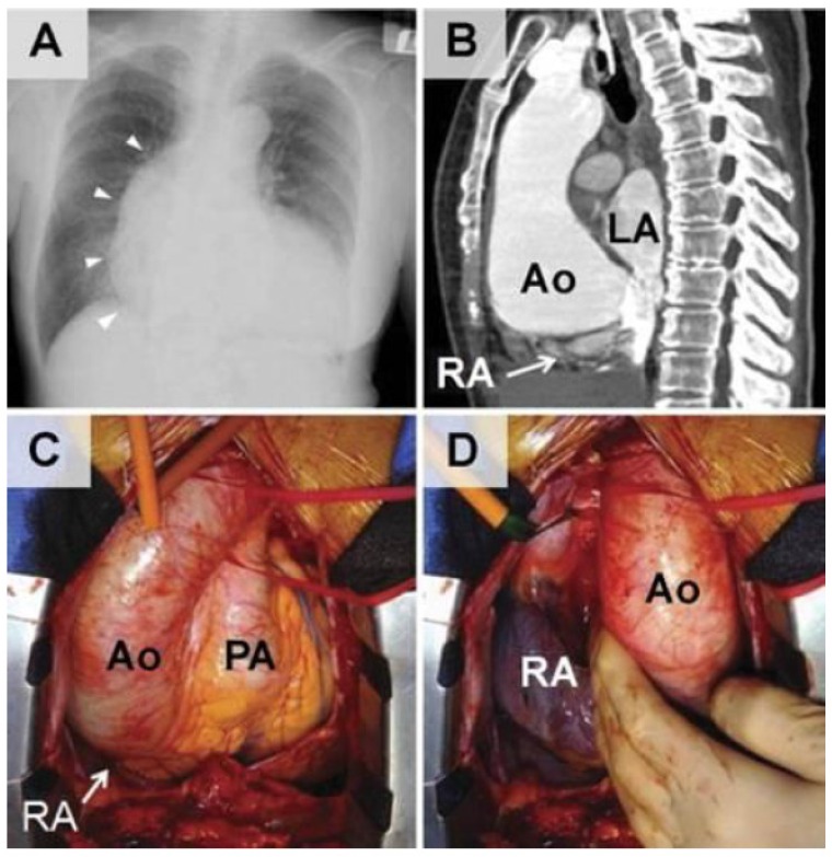
Figure 1.
(A) Chest roentgenogram in the posterior–anterior view reveals pronounced cardiomegaly with marked projection of the right mediastinal border (arrowheads). (B) Sagittal view of contrast-enhanced 64 slice computed tomography shows the enlarged aortic root (Ao), which is posteriorly expanded with compression of the atrial chambers and their septum. (C) In the intraoperative views obtained after sternotomy, the enlarged and elongated aortic root nearly reaches the diaphragm, and (D) manual lifting of the ascending aorta exposes the posteriorly compressed right atrium (RA). (LA = left atrium; PA = pulmonary artery.)

